Laboratoire Matière et Systèmes Complexes ( MSC )
Le laboratoire « Matière et Systèmes Complexes » (MSC) est une unité mixte de recherche du CNRS et de l’université (UMR 7057). Il a pour sujet d’étude la matière et les systèmes complexes sous toutes leurs formes, des fluides montrant des phénomènes complexes non-linéaires aux systèmes proches de la géophysique, de l’environnement et des systèmes vivants.
Agenda
Actualités


Équipes de recherche
DOMM ( Dynamique et Organisation de la Matière Molle )
Auto-organisation et dynamique dans des fluides et composés complexes.
DSHE ( Dynamique des Systèmes Hors Equilibre )
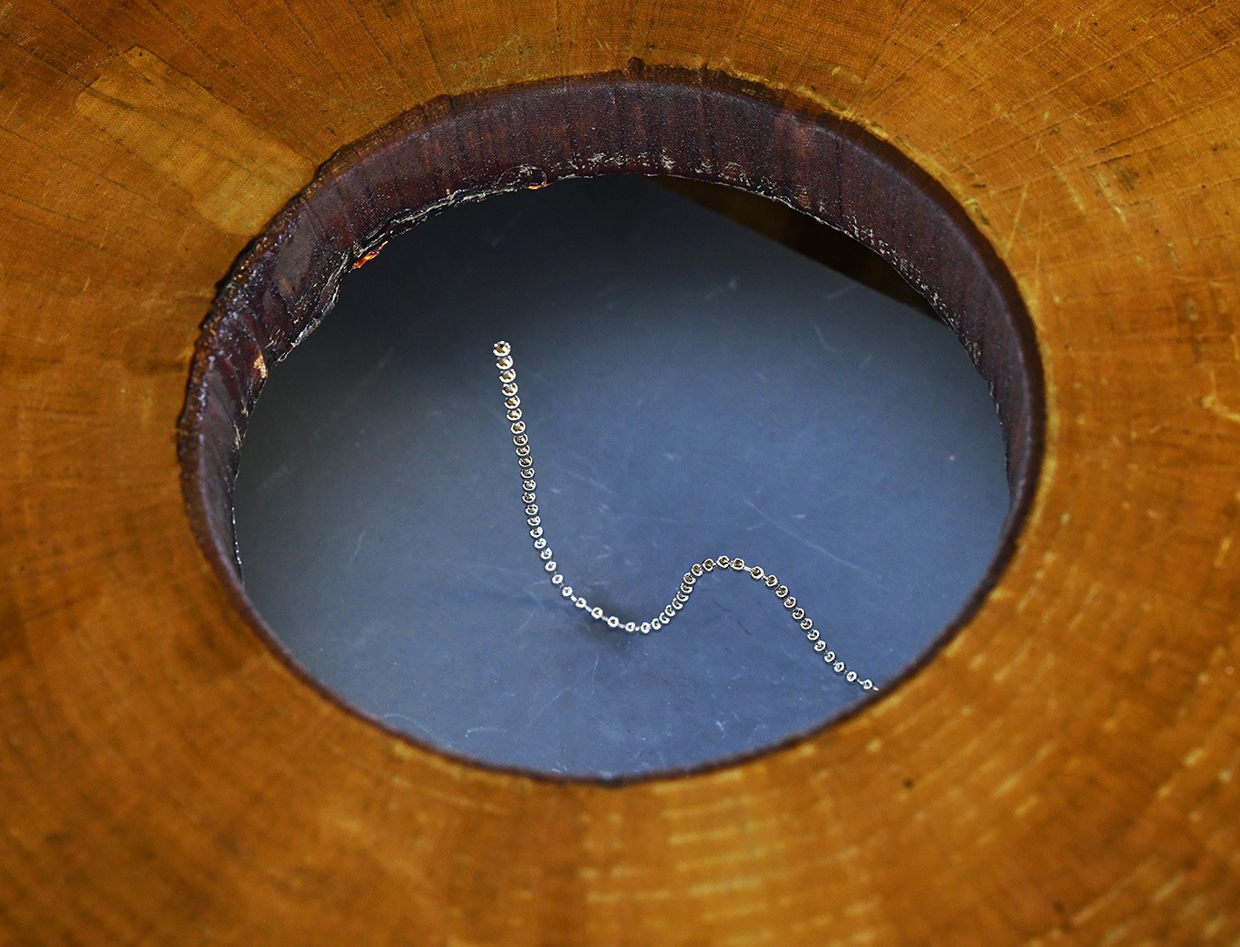
Physique non-linéaire, Hydrodynamique , milieux granulaire, morphogénèse dans l’environnement.
MorphoDyn
Biofluidique, qui s’est encore restructurée en 2023 et a adopté le nouveau nom de MorphoDyn : Morphogenèse et Dynamique des Systèmes Auto-organisés.
MSC-Med
Antenne biomédicale, localisée aux Saint-Pères, Utilisation de nanoparticules ou de vésicules extracellulaires dans un but thérapeutique, Morphogenèse des organes et traitement de leurs anomalies.
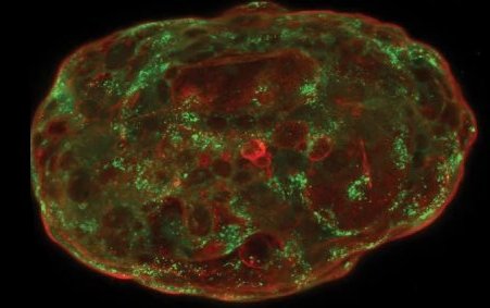
Physique du vivant
Mécanismes Physiques et physico-chimiques du vivant, Mécanobiologie.
Théorie
Physique Statistique Hors Equilibre, Matière Active et Modélisation en Matière Molle et Systèmes Biologiques.
Chiffres clés
Effectif
Permanents
Doctorants Post-doctorants
Visiteurs, CDD
Tutelles


Partenaires

